Imaging and Development Cluster
The cluster of Imaging and Development contains 14 clinicians and scientists from four departments and the School of Rehabilitation Science within the College of Medicine. A general focus of cluster activity is to develop new means to assess and treat skeletal and neural disorders, with a particular emphasis on synchrotron imaging.
By nature, our cluster activities are very interdisciplinary. In addition to having a mix of clinical and basic science members, we collaborate extensively with other disciplines. For example, our focus on synchrotron imaging brings together clinicians, basic scientists, physicists, and engineers. We also collaborate to combine novel imaging techniques with mechanical engineering and 3D-printing tissue engineered constructs, leading the world in non-invasive soft tissue engineering assessments. Our involvement with the nuclear medicine facility further expands this interdisciplinary approach.
|
|
Imaging and Development accomplishments over the past five years have been considerable. We have obtained over 63 grants, 14 of which were co-investigated among cluster members, demonstrating our interdisciplinary interactions. These grants totalled over $30M, with half of cluster members being awarded over $1M the past five years, including 9 CIHR, 12 NSERC, 20 SHRF, and multiple specific disease agency and drug company grants. Highlights of these grants include establishment of the Fedoruk Nuclear Medicine and Imaging Center, a training program for biomedical synchrotron imaging (CIHR-THRUST), a CIHR Synchrotron Medical Imaging grant, and 4 different SHRF Research Team grants (GEMS, TERG, BIG, and OneHealth Imaging). Our cluster has over 193 publications over the past five years, 19 of which were co-authored among cluster members, again illustrating interdisciplinary interactions. 16 more papers are currently submitted. Finally, we have trained at least 140 highly-qualified personnel, including 11 PDF’s, 4 medical residents, 28 PhD’s, 24 MSc’s, 4 medical students, 5 dental students, 39 undergrads, and 26 Research Assistant/Technicians.

Our Research Areas
A general focus of our cluster is on developing new models of skeletal tissue assessment and treatment for skeletal disorders.
Paul Babyn, MD, is the Head of Medical Imaging in CoM and is leading the establishment of nuclear medicine and imaging at the new Cyclotron on campus. He is involved heavily in both clinical and basic science research.
The research program of Soo Kim, PhD, in the School of Physical Therapy broadly encompasses musculoskeletal and shoulder health, using imaging and 3D computer modelling to develop novel ultrasound protocols to assess changes to muscle pre- and post-exercise, treatment, and surgery.
Sheldon Wiebe, MD, MSc, is a clinical radiologist who also specializes in novel synchrotron imaging techniques.
David Leswick, MD is also a clinical radiologist in our cluster.
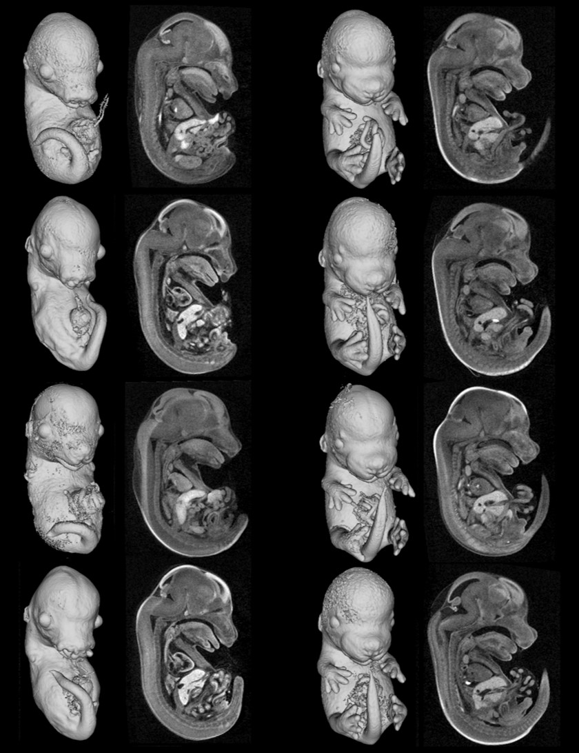
Dean Chapman, PhD, in the Department of Anatomy and Cell Biology is a Canada Research Chair and the current Scientific Director of the CLS synchrotron. His research uses physics to develop novel imaging strategies for both soft and hard tissues, enabling non-invasive assessments of such tricky tissues as lung and cartilage.
David Cooper, PhD, in the Department of Anatomy and Cell Biology is a Canada Research Chair focussed on using synchrotron imaging to track bone adaptation, aging, and disease. He currently directs the BMIT beamline at the CLS, leading efforts worldwide to use synchrotron imaging at doses that enable tracking bone changes longitudinally in the same animal.
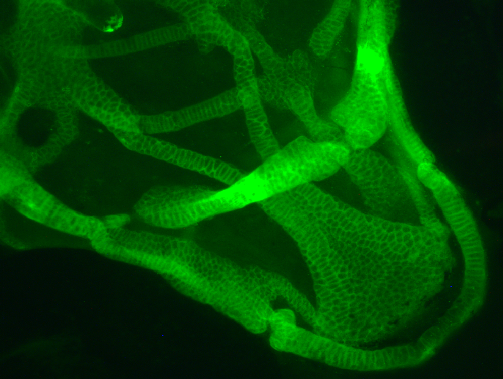
Bill Kulyk, PhD in the Department of Anatomy and Cell Biology studies signalling pathways and extracellular matrix molecules involved in differentiation of embryonic cartilage, applying this knowledge to cartilage tissue engineering approaches.
Julia Boughner, PhD, in the Department of Anatomy and Cell Biology uses synchrotron and other imaging techniques to characterize tooth development and evolution, including the relationship of jaw growth to third molar impactions.
Last, but hopefully not least, Brian Eames, PhD, in the Department of Anatomy and Cell Biology studies the development and evolution of skeletal tissues. Mostly focussed on osteoarthritis, his lab uses zebrafish genetics and synchrotron imaging to understand the molecular mechanisms of osteoarthritis, also developing 3D-bioprinting approaches for articular cartilage tissue engineering.
Another cluster focus is to develop new ways of visualizing and treating neural disorders.
A recent cluster member, Lissa Peeling, MD, in the Department of Surgery, Division of Neurosurgery, works closely with Mike Kelly on a variety of clinical research studies. She recently received a SHRF Establishment Grant to set up her basic research program.
Recent research of Helen Nichol, PhD, in the Department of Anatomy and Cell Biology develops new synchrotron imaging technologies to assess the role of metals in neural disorders, such as Parkinson’s, Multiple Sclerosis, or those resulting from stroke.
Other members of the cluster study basic cellular functions of stress response and metabolism.
Steven Machtaler, PhD
Pat Krone, PhD, in the Department of Anatomy and Cell Biology studies two related subjects: toxicology and stress response. His lab uses synchrotron imaging to relate the localization of toxins (such as mercury) to organ toxicity, and they also study cellular response to heat shock (and toxic) stresses.
Where to Find Us
We are located in the third floor B-wing of the Health Sciences Building.
Health Sciences Building, University of Saskatchewan
107 Wiggins Road
Saskatoon, Saskatchewan, S7N 5E5
Find out more information about Health Sciences at the University of Saskatchewan here.