
Preserving genetic diversity
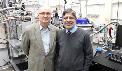
Muhammad Anzar, a research scientist for Agriculture and Agri-Food Canada, is an expert in cryopreservation — you know, freezing living matter to cheat death. His work is more careful than comic book, with a focus not on preserving full organisms but the very germs of life: semen, oocytes and embryos.
“Basically my appointment in Agriculture Agri-Food Canada is about biodiversity and conservation. We are running a Canadian Animal Genetic Resource program to preserve the genetic diversity of our livestock species,” Anzar explains.
In other words, his work is to protect rare livestock breeds from permanent disappearance by preserving their genetic material. In the case of bovine sperm, this is a fairly mundane task, but the female egg, or oocyte, is notoriously hard to preserve.
One of the main goals of cryopreservation is to cool the sample in question with a minimum of ice crystals, which are dangerous since freezing expands and tends to damage the cell’s internal workings. Ice damage varies species to species, animal to animal and even cell to cell, and the reasons for this variation have remained largely mysterious in the field.
That’s why Anzar began work with Canadian Light Source CMCF beamline scientist Pawel Grochulski. CMCF is normally used to study protein structures from crystals, and both scientists thought the technique could be applied to finding troublesome ice crystals in the preserved oocytes.
It worked. For the first time, cryobiology had a way to observe the actual results of the cooling process in a cell.
Now, ideally, instead of forming ice crystals in its cooled state, a properly cryopreserved cell will behave more like a glass — water and any preservatives used will form an amorphous state. The usual technique for this is called vitrification, literally to make a glass.
In vitrification, most of the water in a cell is replaced with cryoprotectants and cooled ultra-rapidly.
“As soon as they are exposed to the high concentrations of cryoprotectants immediately they are plunged into liquid nitrogen with a temperature of about -196C,” said Anzar.
The cells cool at a rate of about 4000 to 5000 C a minute, so ice has almost no time to form, and the cell instead reaches a glass phase.
Instead, CMCF analysis revealed that inside the bovine oocyte, ice crystals still continue to form. Cryoprotectant outside the cells formed glass, as expected, but something about the oocyte is resistant to preservation. In contrast, the bovine embryo was preserved in glass phase efficiently.
Why this is true remains a mystery, but with this new ability to actually observe crystal formation in the vitrification process, Anzar has plenty of plans to continue the exploration.
“The next step might be to use fluorescent markers to tag things important for the cell health, and we’ll be able to study those biomarkers in the frozen state. Or we could look into what is preventing the cryoprotectant to go inside the oocyte.”
“This study opened a gateway to study the behavior of cells at low temperature, and that will be a new dimension for using CMCF to study the frozen cells.”