
ORHG images featured on the cover of Reproduction, Fertility and Development
Ultrasound biomicroscopy imaging allowed non-invasive and repeated monitoring of pig testis cell implants being developed into testis tissues under the back skin of a mouse model.
The work done by Awang Junaidi in Dr. Ali Honaramooz's lab resulted in a front cover image and article in a March 2020 issue of Reproduction, Fertility and Development
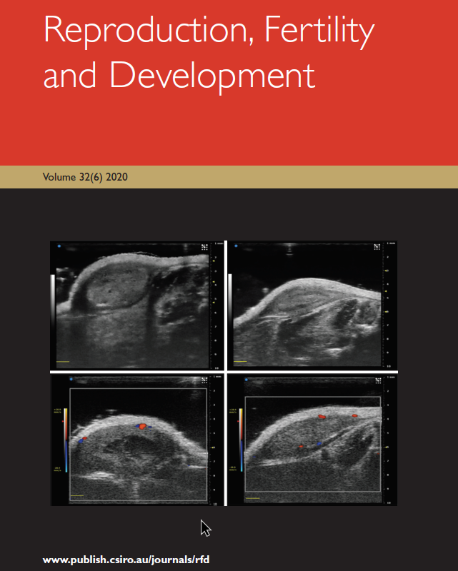
The images highlight the non-invasive application of ultrasound biomicroscopy (UBM) for monitoring the development of testis tissue regeneration, after implantation of pig testis cell aggregates under the back skin of immunodeficient mice. The top row images (using B-mode) show gradual testis tissue formation over time (1 week: top left vs. 2 week: top right), depicted by changes in echogenicity. The bottom row images (using colour Doppler mode) indicate the degree of blood vessel development over time (1 week: bottom left vs. 2 week: bottom right), seen using pseudo-colours (blue; blood flowing towards the probe vs. red: blood flowing away) (Scale bars= 1 mm).