
A day in the life: working in bison reproduction
As the sun rises, Eric and Miranda Zwiefelhofer gear up for another exciting day of work.
By Ella MacQuistenThe husband-and-wife scientists are part of a Western College of Veterinary Medicine (WCVM) research team led by reproduction specialist Dr. Gregg Adams, whose goal is to develop a bison genome biobank — a collection of eggs, sperm and embryos that’s a vital step toward conserving the world’s wild bison population.
While Eric physically collects the oocytes (mammalian eggs) from the female bison, Miranda specializes in maturing and fertilizing the oocytes, and culturing and freezing the resulting embryos — the laboratory work that’s required for in vitro embryo production.
The WCVM’s bison dream team has had many firsts in recent years, and on this particular day, they’re aiming for another first — collecting oocytes from live, pregnant bison and immature bison (pre-pubertal) and using them to create embryos.
Although the University of Saskatchewan (USask) Livestock and Forage Centre of Excellence’s Native Hoofstock Research and Teaching Unit is located just outside of Saskatoon, it’s far enough from the city that visitors get the sense of travelling into the great expanse of the prairies. As we drive past flowering canola fields, Miranda explains the next big step in the road to recovery of threatened bison species.
“To date these oocytes have only been collected from mature non-pregnant bison, which is only a small subset of the wild population,” says Miranda, a PhD student in the USask College of Graduate and Postdoctoral Studies. “Information learned in this study is critical for the next step of the bison genome biobank — which is actually going into the field to collect genetically valuable and isolated genetics of wood and plains bison.”
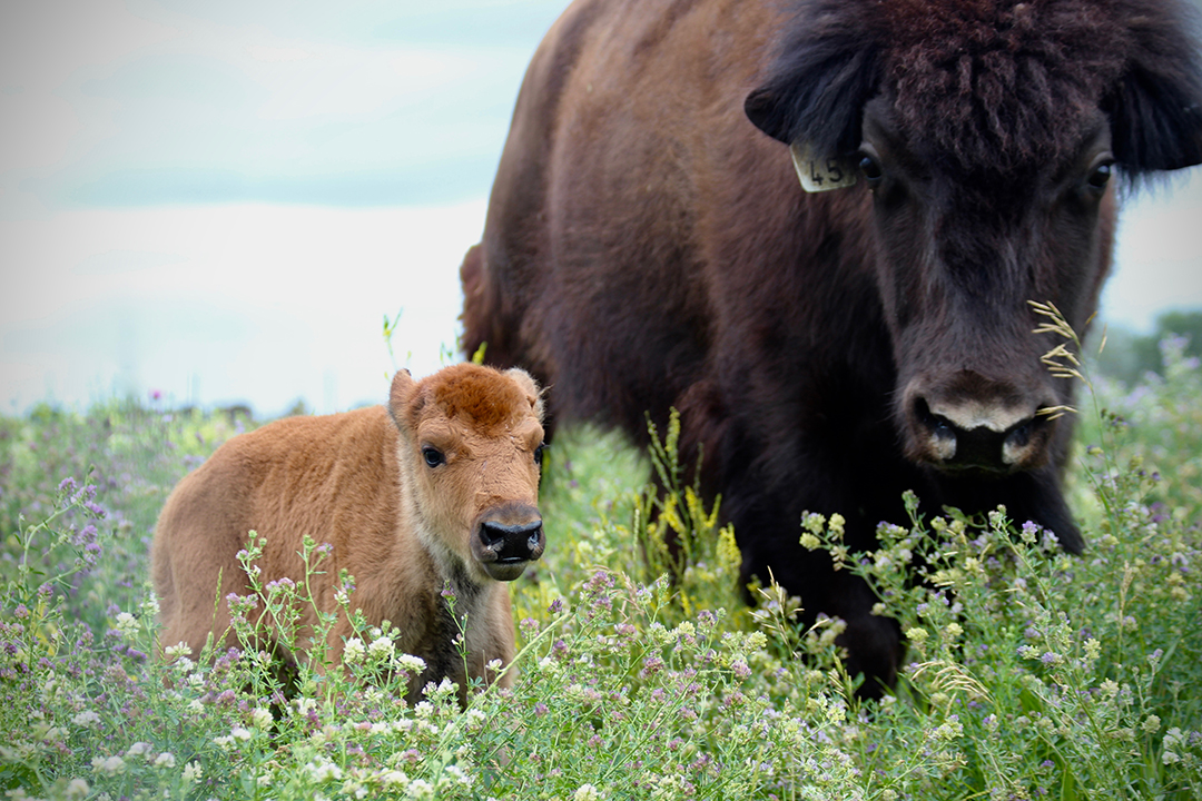
As we approach the farm, the landscape becomes dotted with large, shaggy brunette creatures — the plains and wood bison housed at the centre. They appear to grow in size as we turn onto the narrow gravel driveway.
Once inside the centre’s large barn, Miranda carefully sanitizes and organizes the on-site laboratory in preparation for over 100 bison oocytes, and Eric turns on the hydraulic chute system. The system, which has been specially designed for bison, allows the large animals to move through the barn within high steel walls and various gates without putting the researchers or the bison at risk.
When the team feels set, Miranda climbs to the controls at the top of the hydraulic chute where she has a bird’s-eye view of the bison once they’re inside the barn.
Using immense caution and confidence, Eric herds 12 of North America’s largest land mammals out of their pen and into the barn. They run at an unexpectedly high speed for such large and burly animals, kicking up dust as they approach the barn.
Once the first bison cow is secured in the chute, the real work begins. Eric pops open a small door near the rear end of the bison and administers an epidural (local anesthetic drug) so that “the bison does not feel a thing.” It’s an important step that makes it easier for him to manipulate the reproductive tract.
“The goal of follicular aspiration is to collect the oocytes from the follicles on the ovary,” explains Eric as he puts on a long plastic sleeve that covers his entire arm.
With the ultrasonography machine at his side, Eric begins the oocyte collection procedure, which is technically known as “transvaginal ultrasound-guided oocyte retrieval.” Eric breaks down the steps of the procedure as he works.
“The transvaginal ultrasound probe is gently inserted into the vagina. Once the probe is inserted, I place my left hand into the rectum and remove as much feces as I can,” says Eric as he applies lubricant to his gloved hand and begins shovelling out feces.
It’s not a glamorous job, but it is one that Eric knows well.
“I grew up on a small dairy farm in Wisconsin, so I have worked with cattle my entire life,” says Eric, a reproduction specialist and a postdoctoral researcher with USask and the Toronto Zoo. “Bison and cattle both exhibit a similar reproductive tract, so the techniques are similar between the two species.”
As he continues the process, Eric explains the next steps: “I can begin to carefully palpate the reproductive organs such as the cervix, uterus and ovaries. I bring the ovaries toward the ultrasound probe, and I can visualize the ovarian structures on the ultrasound screen.”
As he works, a round grey structure containing multiple black circles appears on the screen. Those black circles are the follicles in the ovary, and each follicle contains an oocyte. While the imaging allows Eric to guide the needle into the follicle, his assistant switches on the vacuum pump connected to the needle and the black circle on the screen shrinks and disappears.
The tiny oocyte has been moved from the follicle within the ovary to a container that’s encased by a mini portable incubator — temperature is just one of many factors that needs to be regulated in order to ensure the survival of these delicate oocytes.
Working with wildlife — especially herding and handling bison — is no easy feat, but Miranda and Eric work with ease and confidence after much practice.
“Just simple activities such as moving bison can be a challenge, so you always have to be thinking when you are around them and show them the respect they deserve,” says Eric.
The entire process from catching the bison to release takes about 15 to 20 minutes, but it’s important that the process be as stress-free and as fast as possible for the animal. After Eric is done, he releases the squeeze on the bison cow, and it exits back into the sunny pasture. The meticulous process is then repeated for the next animal.
Once the collected oocytes and follicular fluid have been carefully carried to Miranda in the lab, it’s her turn to take care of the oocytes. She filters the fluid and uses stereomicroscopy — a special microscope that produces three-dimensional images — to count the oocytes before methodically placing them in a special medium that will promote their maturation.
Although the entire process is painstaking at times, Miranda says she’s reminded of how rewarding their work is every time she drives out to the farm.
“The past two summers we have had bison calves born from embryos that took countless hours at the farm and in the lab to produce,” says Miranda. “It is very rewarding to see them out in their pastures. Each time I see my calves, I feel like all of the hard work was worth it.”
After a long day at the farm, Eric and Miranda load the collected oocytes into the car and head back to the USask campus and the Westgen Research Suite at the WCVM — a laboratory devoted to advanced reproduction techniques. Once there, the oocytes will mature before undergoing in vitro fertilization with sperm.
“The most gratifying part of the project is that we can apply these techniques that we have developed and may be able to save a threatened species,” says Eric.
“It is very rewarding when we can look out into a pasture and see a newborn calf born from our methods and we can say to ourselves, ‘We helped to make that calf.’”
Ella Macquisten of Victoria, B.C., is a second-year veterinary student at the Western College of Veterinary Medicine (WCVM) whose research position was supported by the college’s Interprovincial Undergraduate Student Summer Research program. Her story is part of a series of articles written by WCVM summer research students.