Research
Interests
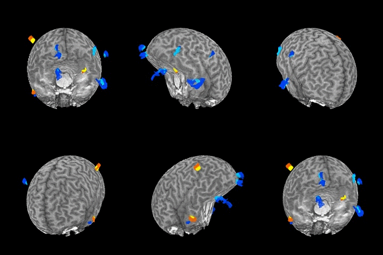
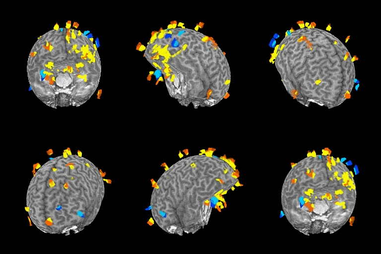
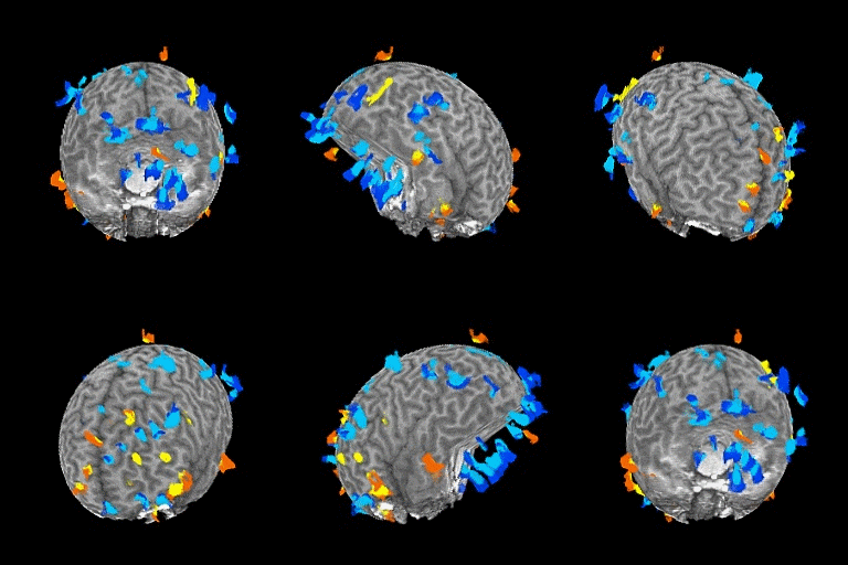
- Neuroimaging (functional Magnetic Resonance Imaging, fMRI; Diffusion Tensor Imaging, DTI)
- Mental chronometry (reaction time, response duration, accuracy)
- Connecting the former to the latter (i.e., the brain to the mind) in language, semantic, attention, memory and object processing networks through computational modelling
- Ventral and dorsal visual processing streams
- Embodied cognition (sensorimotor-semantic processing involved in language)
- Reading systems
- Localization of these networks in pre-neurosurgical planning


Below is a video of a Diffusion Tensor Imaging (DTI) map, which reflects white matter tracts (glial cells and myelinated axons) that transmit signals between brain regions. Our lab uses both fMRI and DTI techniques in basic research and neurosurgical assessment "from bench to bedside and back again".
Below is the 2 minute segment describing our lab's research as it relates to "Thinking" (part of Collaboration Collider where we brought together researchers from across the College of Arts & Science, as part of a Science Meets Arts Plus Public "SMART+P" project).
Recent virtual conferences
Our lab presented research at the virtual conferences for the Cognitive Neuroscience Society (CNS), the Organization for Human Brain Mapping (OHBM), and the Society for Neurobiology of Language (SNL). Here are links to some of our poster presentations: Neudorf et al. CNS, OHBM; and Kress et al. OHBM, SNL.
