Primary Investigator: Dr. Michael Kelly
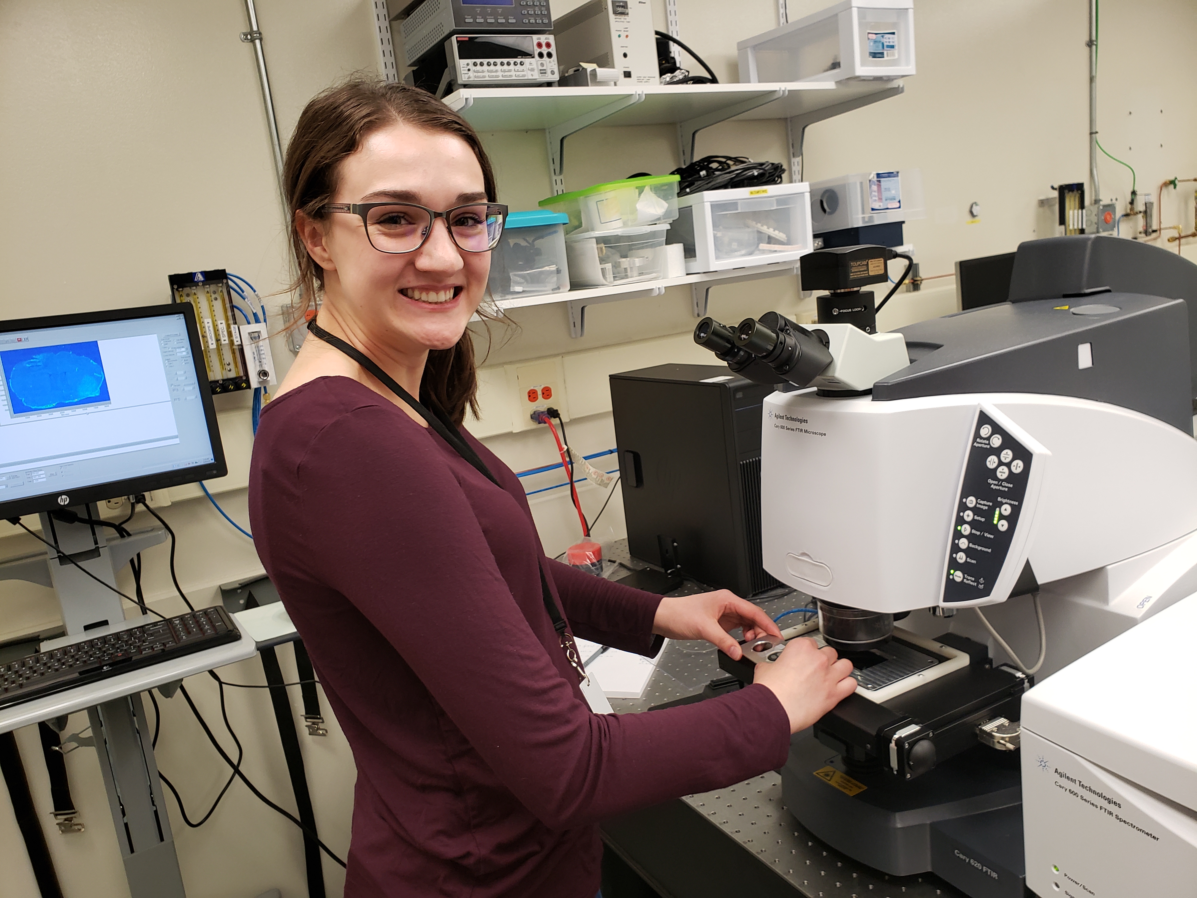
To better understand the mechanisms of cellular damage following a stroke the Kelly lab has developed a novel imaging approach, comprised of experts in stroke research and synchrotron imaging. The team combines synchrotron-based X-ray fluorescence imaging (XFI) with Fourier transform infrared (FTIR) spectroscopic imaging as well as conventional methods, including histology and immunohistochemistry, to reveal new knowledge about changes in the brain after stroke. Our imaging approach also affords a method for differentiating the penumbra, a region of tissue surrounding the core of the stroke lesion which has not yet died and thus represents a critical clinical target for therapeutic intervention.

