Saskatchewan Cerebrovascular Centre Research
Clinical Stroke Research
The SCVC is actively participating in 18 clinical studies, with another 6 studies in start-up. We have previously completed participation in 31 additional clinical studies to date, all to help focus our efforts on delivering the best patient care.
Retrospective Analysis
The SCVC is currently participating in one retrospective analysis trial, with completed participation in 6 additional trials.
Registries
The SCVC is currently participating in 4 registries, with completed participation in an additional 10 trials.
Drug Trials
The SCVC is currently participating in 9 drug trials, with completed participation in 6 additional trials.
Device Trials
The SCVC is currently participating in 2 drug trials, with completed participation in 2 additional trials.
Treatment Trials
The SCVC is currently participating in 3 drug trials, with completed participation in 7 additional trials.

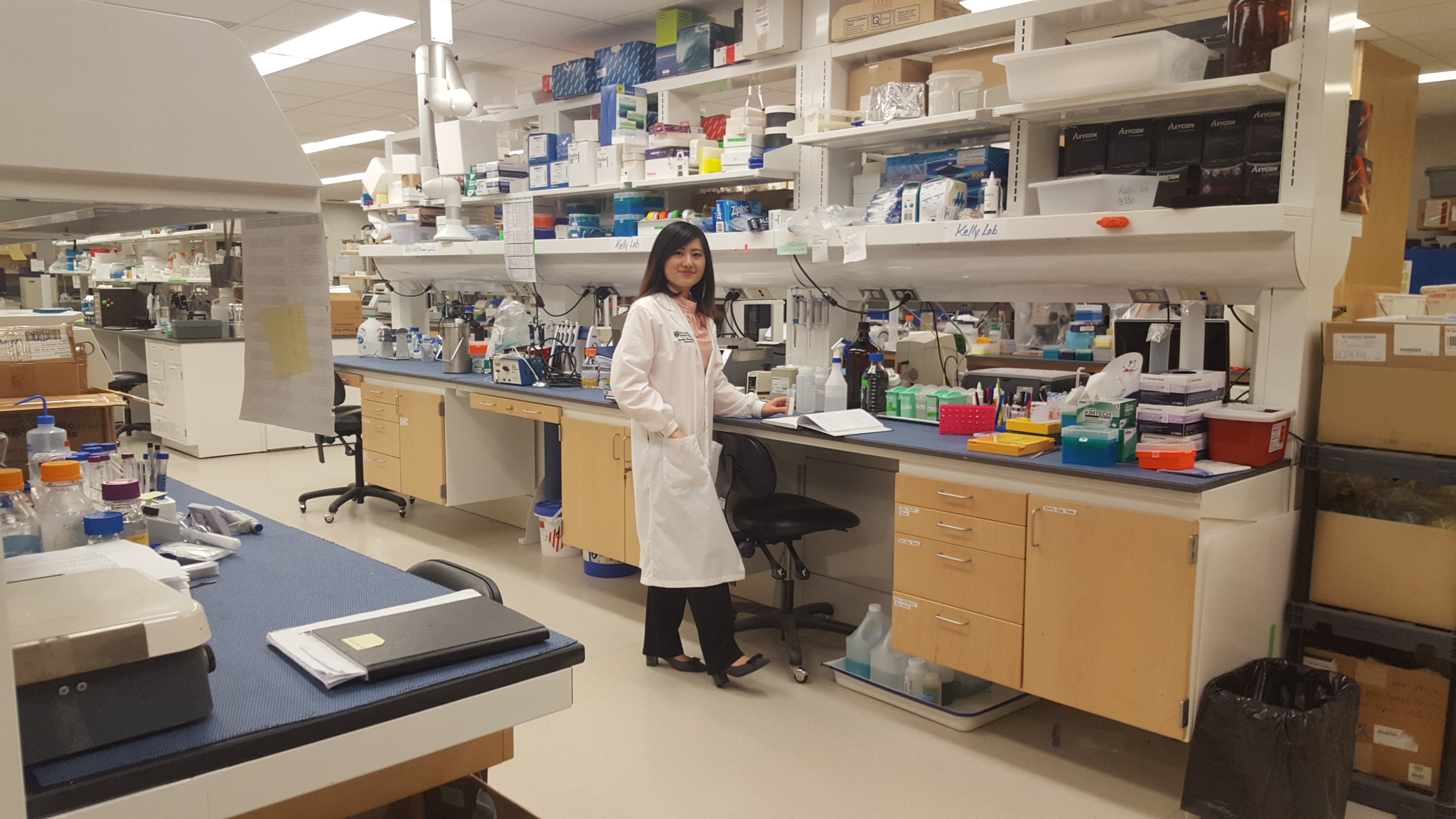
Bench Science Research
Bench science stroke research is performed using a collaborative approach. Director, Dr. Michael Kelly, is a member of the HR/HSFC Team in synchrotron medical imaging. The research currently has three main focuses: brain metal changes after stroke; studying neuroendovascular devices in animal models; and the evaluation of computational flow dynamics within a side-wall aneurysm model using the synchrotron. Synchrotron imaging techniques such as diffraction enhanced imaging (DEI), rapid scanning X-Ray diffraction and K-edge digital subtraction angiography (KEDSA) are being used and bioabsorbable stent studies are currently in development.
Currently, the bench science research led by the SCVC in collaboration with others, has examined the time course of biochemical changes that surround brain tissue damaged by stroke. The specific focus of this research has been to identify the biochemical mechanisms that drive the spread of tissue damage from the initial stroke lesion into healthy tissue. The use of the novel biochemical imaging techniques in the project has been applied to a mouse model of ischemic stroke and has enabled biochemical characterization of tissue that is “at risk” of further damage following ischemic stroke.
Animal Models of Ischemic Stroke
Enhancing our knowledge of the underlying biochemistry and cellular mechanisms of stroke for the development of innovative therapies to reduce the tremendous burden that stroke has on society.
Metabolic and Biomolecular Imaging
Our lab has developed a novel immaging approach, comprised of experts in stroke research and synchrotron imaging, to better understand the mechanisms of cellular damage following a stroke.
Following Metabolic Markers
Research investigating the usefulness of Trifluoperazine (TFP) and hypothermia to reduce the amount of swelling and cell death in the brain tissue surrounding the stroke lesion.
Imaging Biomarkers of Hemmorhagic Transformation
We have identified a range of metabolic biomarkers from X-ray fluorescence imaging, X-ray absorption spectroscopy, and Fourier transform infrared imaging that are highly correlated with blood components.
Mouse Models of Intracerebral Hemorrhage
Dr. Peeling’s research aims to establish a mouse model of intercerebral hemorrhage (ICH) at the University of Saskatchewan, using collagenase injection.
Aneursym Blood Flow Dynamics
Assessment of a novel in vitro approach to quantitatively and qualitatively assess flow dynamics of porcine red blood cells within a side-wall aneurysm.

