
Access to Equipment
To arrange access to the Cancer Cluster instruments please contact:
 |
Mark Boyd Lab Manager of 4th floor, and 6th Floor Office Room 4B30 Phone: (306) 370-1296 E-mail: mark.boyd@usask.ca |
See Expert User Database for contact information for an expert user that can provide the necessary training needed to use the equipment for the particular application needed.
If the data obtained using the Cancer Cluster instrumentation are used for publication, please include the statement below in the acknowledgements section:
"Research described in this paper was performed in part at the Cancer Research Cluster, which is supported by the Saskatchewan Cancer Agency and the College of Medicine, University of Saskatchewan."
Bacterial Shakers
Three Bookable Bacterial Shakers
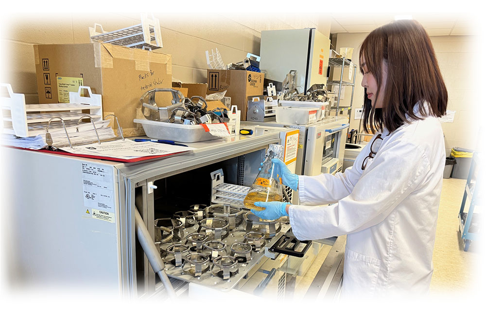
Adjustable temperatures, including cooler than room temperture.
Adjustable shaking speed.
Capable of holding many difference sized flasks, tubes and/or plates with appropriate clamps.
Confocal Microscope
Zeiss LSM900 Airyscan 2 system
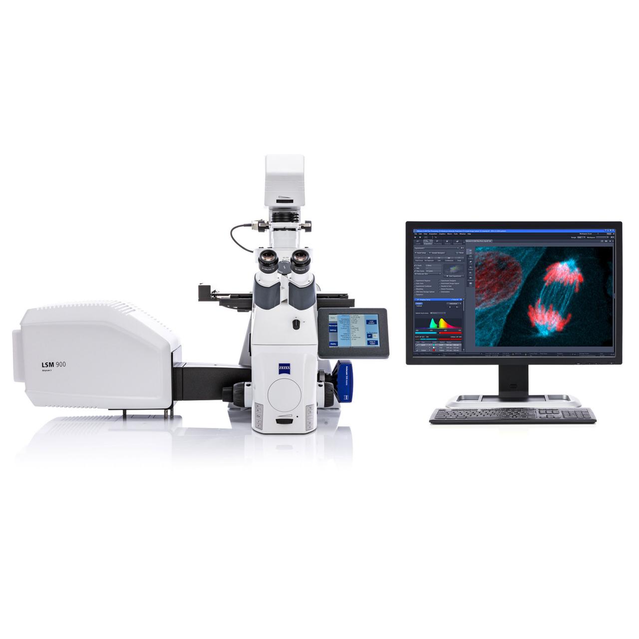
- ***NEW - Will be available for free training starting Dec 12, 2024 and then booking to use
- User fees apply, $40/hr; Note billing will be based on the data on Q-Reserve to your on file CFOPAL by JV
- Click here to view booking calendar in Q-reserve.
- immunofluorescence microscopy focused on one 'layer' of the cell
- Objectives: 2.5x, 10x, 20x, 40x/NA1.2 water immersion, 66x/NA1.4 oil immersion
- can build 3-D images by taking multiple Z-stacks
- coimmunofluorescence and colocalization using software analysis
- fixed or live cells imaging
- video imaging capability to capture imaging over time
- multiple fluores can be used in the same sample and the same time
- GFP. RFP, CFP. YFP, DAPI, brightfield
- Alexa dyes, Cy dyes
- spectral unmixing capabilities, even for dyes with overlapping spectra
- FRET, FRAP experiments
- significant training required
- See product info
Purchased with funds from the Saskatchewan Cancer Agency.
![]()
Flow Cytometer
Beckman CytoFLEX Flow Cytometer
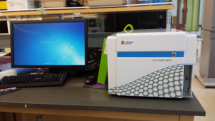
- View Booking Calendar here
- User fees apply, $50/hr
- 3 lasers; 488 nm, 638 nm, 405 nm
- capable of detecting 13 colors at once
- 96-plates or single samples
- Quantification of cell surface markers or intracellular proteins
- Requires antibodies to target protein
- Cell cycle analysis
- Apoptosis assay
- More info here: https://www.beckman.com/flow-cytometry/instruments/cytoflex
Purchased and maintained with funds from the SCA ![]()
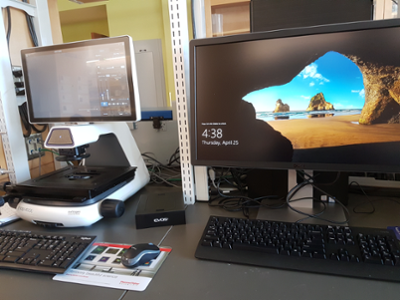
Fluorescent Microscope System (EVOS 5000)
EVOS 5000 and Celleste Analysis Software workstation
- View Booking Calendar here
- User-defined fluorescence, transmitted light, and color applications
- Autofocus, Z-stack capability, time-lapse imaging, and single-click multi-channel capture
- On-board software for acquisition, analysis, and automated cell counting
- Light cubes: DAPI, GFP, TX RED
- Objectives: 4X, 10X, 20X, 40X, 60X
- Wide variety of sample holders for slides and various sizes of plates/dishes
Celleste Image Analysis software available to use on-site at computer workstation beside EVOS5000.
Purchased and maintained with funds from the SCA ![]()
Fluorescence Microscope System (Olympus IX51)
Olympus IX51
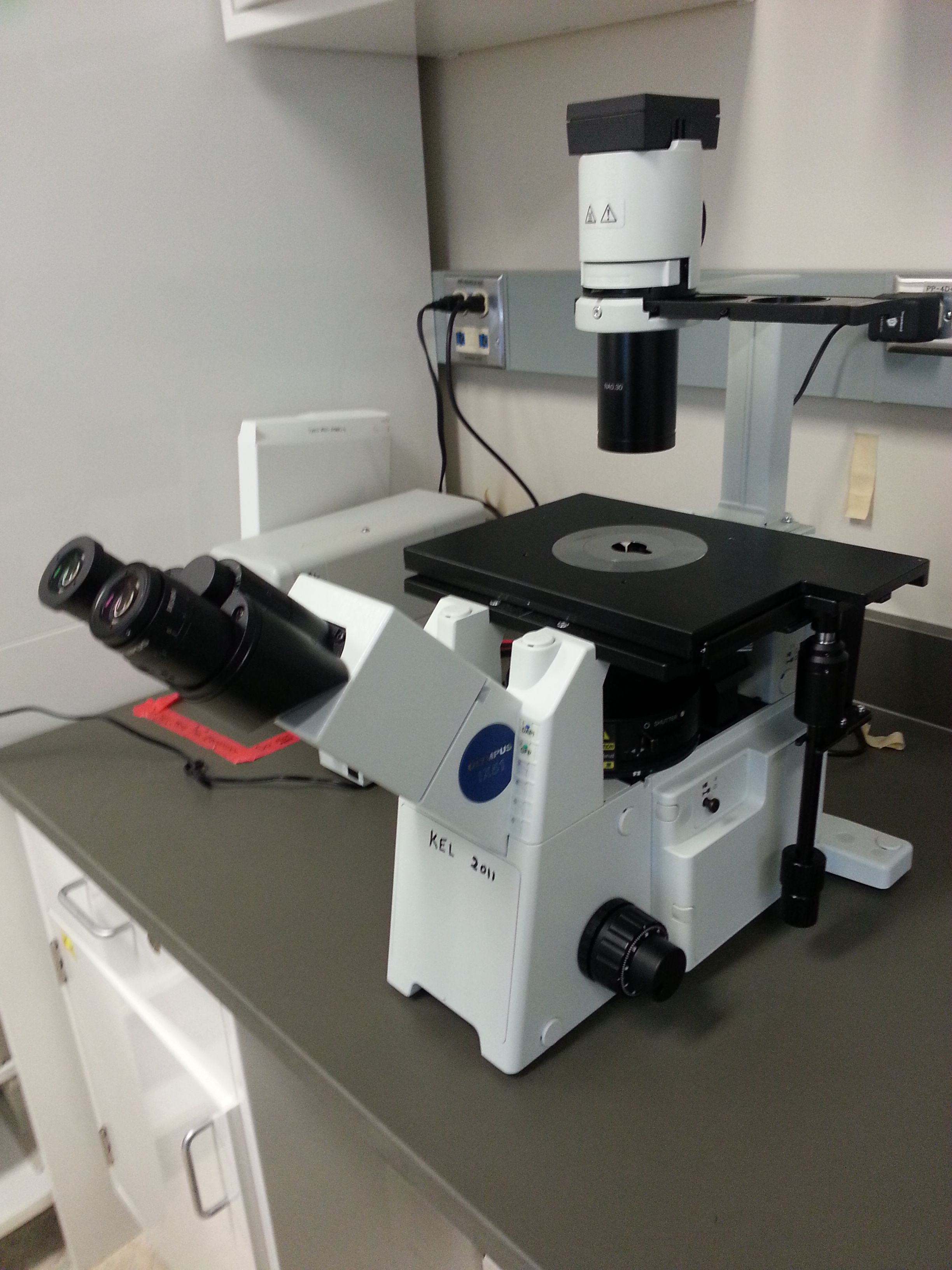
- immunofluorescence microscopy
- green, red, blue, brightfield
- e.g. GFP, RFP, DAPI, light images
- photography capability
- permission of owner Dr. Lukong required
Hypoxia Chamber
BioSperix - ProOx Model 110
- senses and displaces oxygen on an ongoing basis
- requires user supplied nitrogen tank
- oxygen gas sensor can be set to maintain desired oxygen level
- fits inside existing CO2 incubator
Purchased with funds from the SCA ![]()
Imaging System - BioRad
ChemiDoc MP Imaging System - 17001402
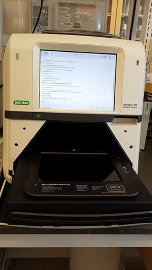
- View Booking Calendar here
- multiplex fluorescent western blotting
- chemiluminescence detection
- general gel documentation applications and stain-free technology
- fluorescent reporters (RGB, far red, and near infrared) including StarBright™ Fluorophores
- detect up to three proteins simultaneously
- features include automatic selection of optimal light source by application, auto focus, auto exposure, and preview features ensure optimal images
- stain-free imaging permits the normalization of bands to total protein in both in gels and blots; eliminates the need for housekeeping proteins
- three sample trays available to cover diverse imaging applications. Smart Tray Technology automatically recognizes the application-specific trays and adjusts imaging parameters and software options accordingly
- White Sample Tray, for gels stained with Coomassie Blue, copper, silver, or zinc stains
- Trans-UV, 302 nm
Epi-white
Trans-white (White Sample Tray required)
Trans-blue, 450-490 nm
Epi-blue, 460-490 nm excitation
Epi-green, 520-545 nm excitation
Epi-red, 625-650 nm excitation
Epi-far red, 650-675 nm excitation; so suitable for IR-680 dyes (like LICOR)
Epi-near IR, 755-777 nm excitation; so suitable for IR-800 dyes (like LICOR)
Note: an identical BioRad Chemidoc system is also available to use and is located in the Common Equipment Core Facility in B128, HSc here.
Purchased with funds from the SCA ![]()
Imaging System - not chemi
Axygen gel doc
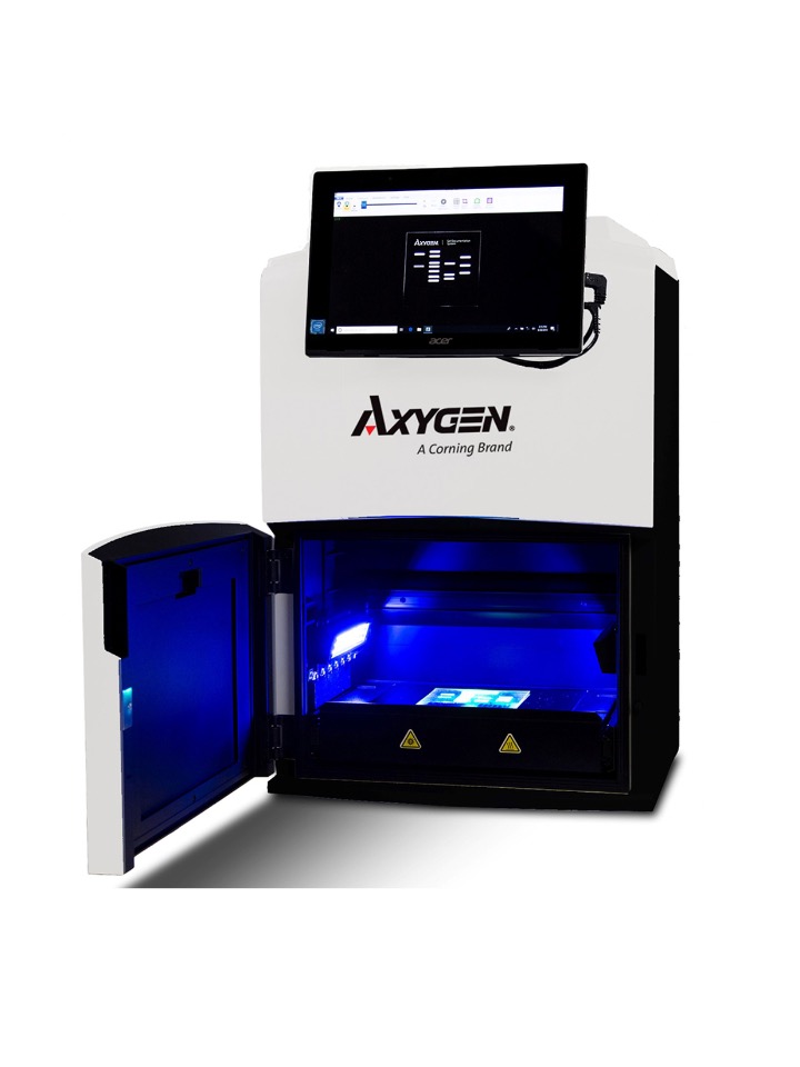
- Axygen® Gel Documentation systems easily capture publication quality, 16 bit TIFF images.
- Darkroom tab lets you select from UV 302, UV 365, Epi White, or Epi Blue (BL system only) light sources, with a Trans White light illumination tray.
- Auto Exposure tool calculates the optimal exposure time with just a single click. You can also use the Slider tool to manually adjust the exposure time and view an updated live image.
- Select ROI tool allows you to select a region of interest. The Auto Exposure calculations are only based on the selected region. This feature is especially useful for gels that have either intense or faint bands.
- Camera: 5.4 MP image resolution
- Lens: f1.8
- Light Sources: • Epi White; • Dual wavelength transillumination at 302 nm and 365 nm; • Epi Blue
- Trans White Light Illumination Tray: We have this too
This system is best suited for quick gel image capture of DNA and protein gels and does NOT have chemilluminescence features. There is no sign up, since use is quick.
A thermal printer is on order.
Imaging Live Animals - IVIS Spectrum
The IVIS® Spectrum in vivo imaging system combines 2D optical and 3D optical tomography in one platform. The system uses leading optical technology for preclinical imaging research and development ideal for non-invasive longitudinal monitoring of disease progression, cell trafficking and gene expression patterns in living animals.
Features and Benefits
- High-sensitivity in vivo imaging of fluorescence and bioluminescence
- High throughput (5 mice) with 23 cm field of view
- High resolution (to 20 microns) with 3.9 cm field of view
- Twenty eight high efficiency filters spanning 430 – 850 nm
- Supports spectral unmixing applications
- Ideal for distinguishing multiple bioluminescent and fluorescent reporters
- Optical switch in the fluorescence illumination path allows reflection-mode or transmission-mode illumination
- 3D diffuse tomographic reconstruction for both fluorescence and bioluminescence
The equipment is located in the Health Sciences Vivarium. The receive training and arrange access, please contact Michele Moroz (michele.moroz@usask.ca).
Incucyte S3 Live Cell Analysis System
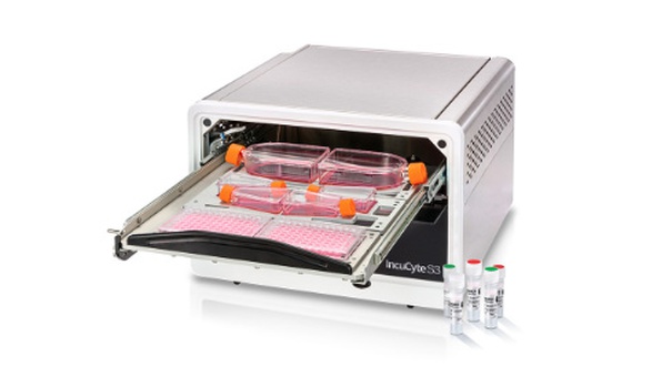
- ***NEW - Starting Jan 1, 2025 user fees will be charged for use
- User fees apply, $10/day; Note billing will be based on the data on Q-Reserve to your on file CFOPAL by JV
- View booking calendar here in Q-reserve.
- Live cell imaging e.g., proliferation, or cytotoxicity assays
- Leave your cells undisturbed in the incubator during image acquisition
- Multiple imaging modes – HD phase plus red and green fluorescence
- 4x, 10x and 20x objectives
- Accommodates up to six microplates at a time
- Users can schedule experiments at different image acquisition frequencies and magnifications in parallel
- Remote, networked access with unlimited, free licenses
- Incucyte compatible vessels can be found here
- Incucyte handbook can be found here
Purchased with funds from the Cancer Foundation of Saskatchewan. Maintained with funds from the SCA.
![]()
LICOR CLx Odyssey Scanner
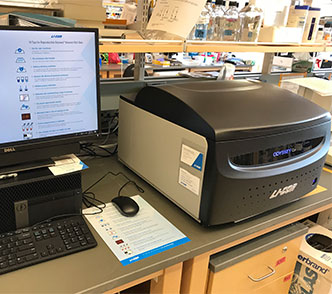
- View Booking Calendar here
- quantitative Western/Immunoblots for protein detection
- much less expensive than chemiluminescence (no film, no chemi solutions)
- uses infrared-linked secondary antibodies (very economical)
- directly scans blots to provide accurate quantification over several orders of magnitude
- more sensitive detection than film (from chemi)
- digital image of same quality as film (from chemi)
- two channels to detect two species of antibodies at the same time (e.g. mouse and rabbit)
- in-cell Western to carry out higher throughput analyses in 96-well format for validated antibodies that only recognize one protein in cells
Note: an similar LICOR Odyssey scanner system is also available to use and is located in the Common Equipment Core Facility in B128, HSc here.
Purchased and maintained with funds from the SCA ![]()
Luminometer
GloMax(R) 96 Microplate Luminometer w/ Dual Injectors from Promega
- View Booking Calendar here
- chemiluminescent and bioluminescent applications
- highly sensitive and quantitative; nine logs of dynamic range
- capable of performing both flash and glow-type luminescent assays
- injector volume range from 25 - 250 ul in 1-ul increments
- software wizards assist with protocol development
- Suitable for: dual reporter assays, steady-Glo® Luciferase Assays, Flash Glow Luciferase Assays, Cell Viability/ATP Assays, Kinetic Assays, intracellular Ca+2 Assays, Immunoassays
- Spectral Range: 350-650 nm
- Detection Limit: 3 x 10-21 moles of luciferase
- Requires the use of 96-well microplates
- Data exports into Excel spreadsheet
Purchased with funds from the SCA ![]()
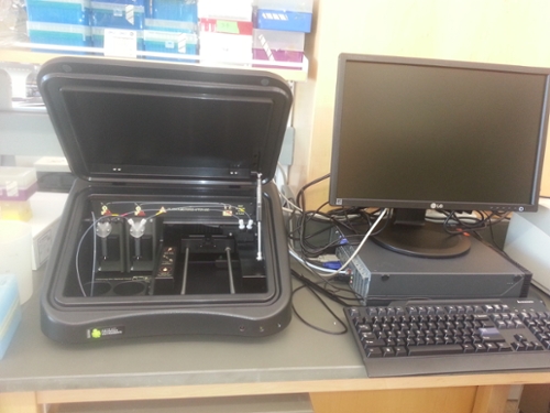
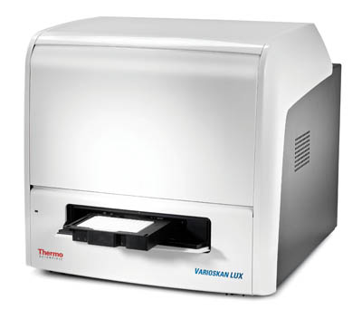
Microplate Reader
Varioskan Lux Multimode Microplate Reader
- View Booking Calendar here
- Absorbance (UV-Vis, including pathlength correction)
- Fluorescence Intensity (including FRET)
- Luminescence (direct and filtered, including BRET)
- AlphaScreen / AlphaLISA
-
The instrument selects the measurement wavelength using filters or monochromators, depending on which is optimal for each measurement technology.
- Monochromators in absorbance and fluorescence intensity
- Filters in AlphaScreen and time-resolved fluorescence
- Luminescence without wavelength selection (filters can be used if required)
- Absorbance wavelength range: 200 - 1000 nm
- Light Source: Xenon Flash Lamp
- Fluorescence excitation wavelength range: 200 - 1000 nm
- Fluorescence emission wavelength range: 270 - 840 nm
- Luminescence wavelength range: 360 - 670 nm
- Measurment modes: End-point, kinetic, spectra, multipoint and kinetic spectra
Purchased and maintained with funds from the SCA ![]()
Nanodrop
Nanodrop 2000C
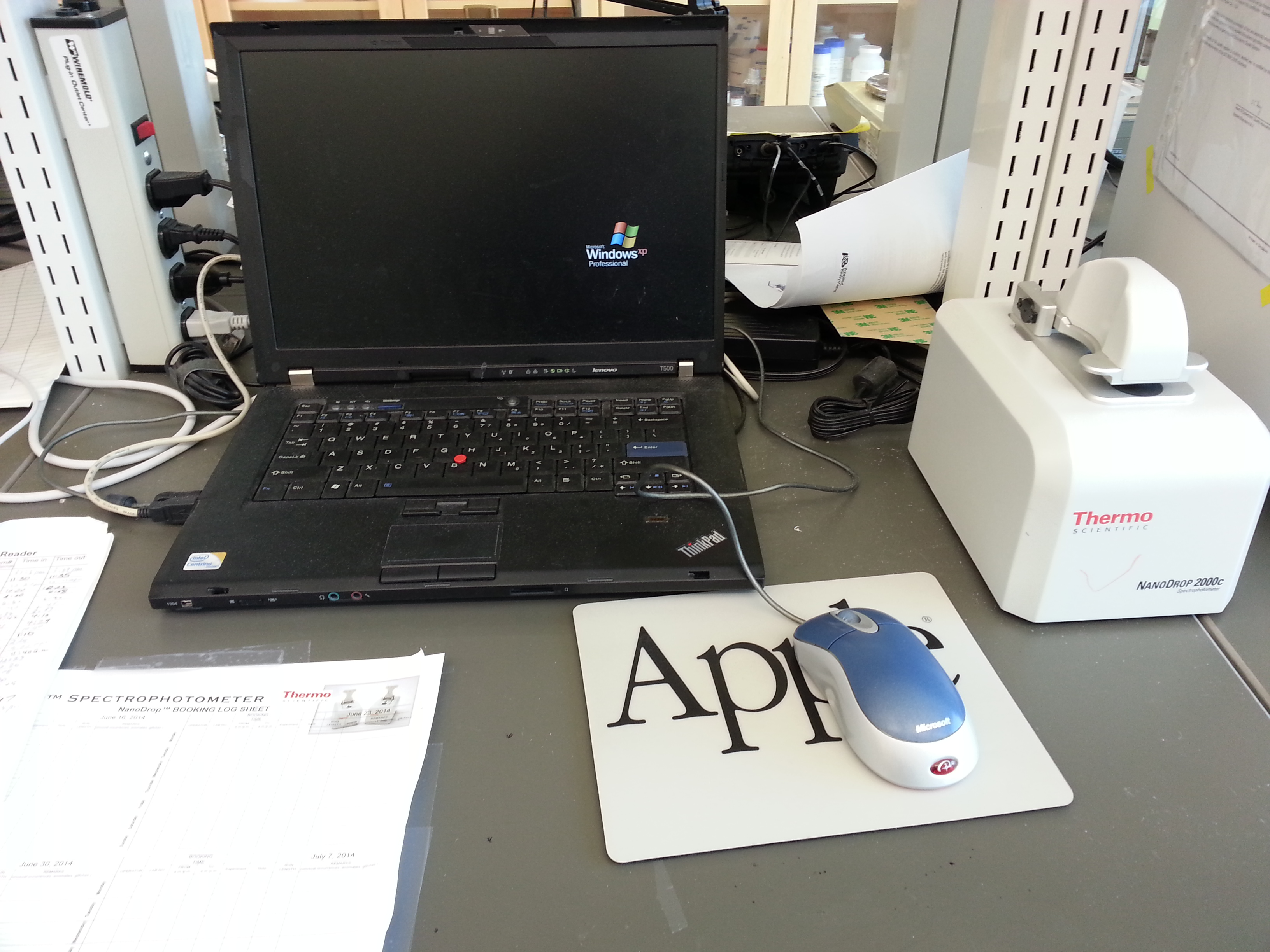
- View Booking Calendar here
- determination of protein or nucleic acid concentrations
- uses very small sample sizes
Purchased with funds from the SCA ![]()
Next-Generation Sequencing Facility
The Next-Generation Sequencing Facility (NGSF) is a high-throughput genomics facility located at the University of Saskatchewan. It is supported by the Saskatchewan Cancer Agency and University of Saskatchewan, College of Medicine. The primary goal of the NGSF is to further basic and clinical research by providing access to high-throughput sequencing technologies and expertise.
In addition to library preparation and sequencing services, data analysis and bioinformatics services are also available, either together or each independently.
Visit the main NGSF page for more information, including a list of shared equipment (Qubit, Tapestation, qPCR, iSeq, NextSeq and more). Contact us to discuss your project objectives and obtain a service quote.
![]()
![]()
Phenogenomic Imaging Centre of Saskatchewan
- gene knockdown (shRNA) and gene knockout (CRISPR) technology
- high-content screening and imaging capabilities
- User fees apply
- ImageXpress Micro XLS Widefield High Content Screening System
- ImageXpress Ultra Confocal High-Content Analysis System
- Capable of fluorescence imaging of 96-well plates
- Pooled shRNA library, lentiviral delivery
- Arrayed shRNA library, lentiviral delivery
- CRISPR-Cas9 gene editing tehcnology


Phosphorimager
Typhoon FLA 7000, from GE Healthcare
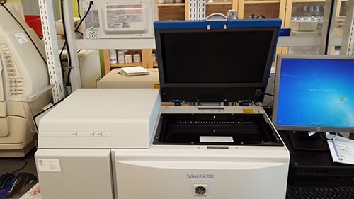
- View Booking Calendar here
- Typhoon FLA7000 Scanner Main Bi-Alkali PMT Laser
LD 635 Laser LD 650 Laser LD 473 Laser SHG 532 Y520 filter (FLA7k) O580 filter (FLA7k) R670
filter (FLA7k) IP filter (FLA7k) Fluor stage (FLA7k) Membrane weight (FLA7k) Phosphor stage
(FLA7k) Fluorescent plate Capture software - digital detection and quantification of 32P and 35S-based radioactive assays on gels and/or TLC plates; requires high resolution phosphor screens to capture information. Some shared and available for use from Deborah Anderson (deborah.anderson@saskcancer.ca)
- Fluorescent gel imaging of DNA and Protein
- Fluorescent Western blot imaging
Purchased with funds from the SCA ![]()
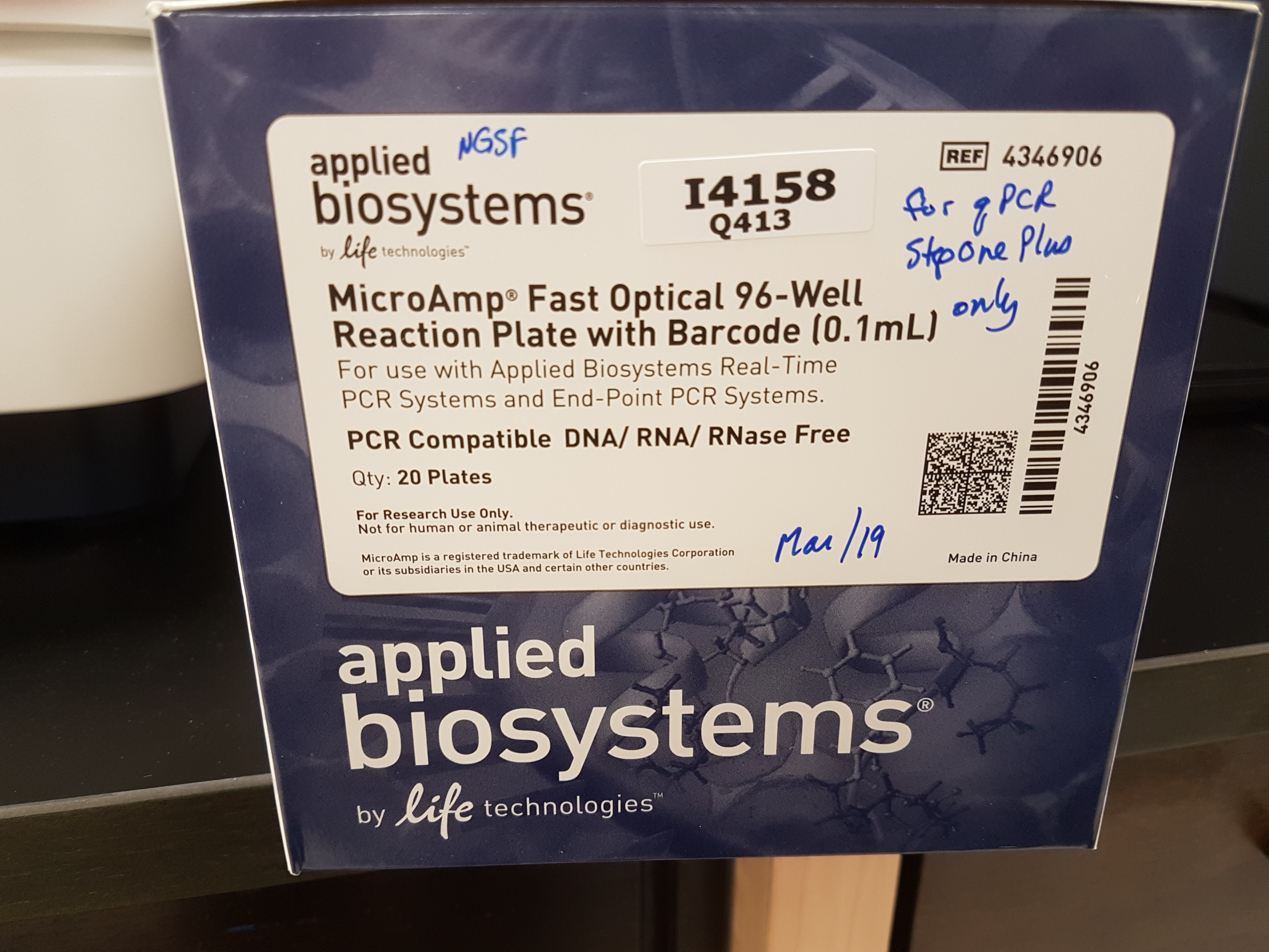
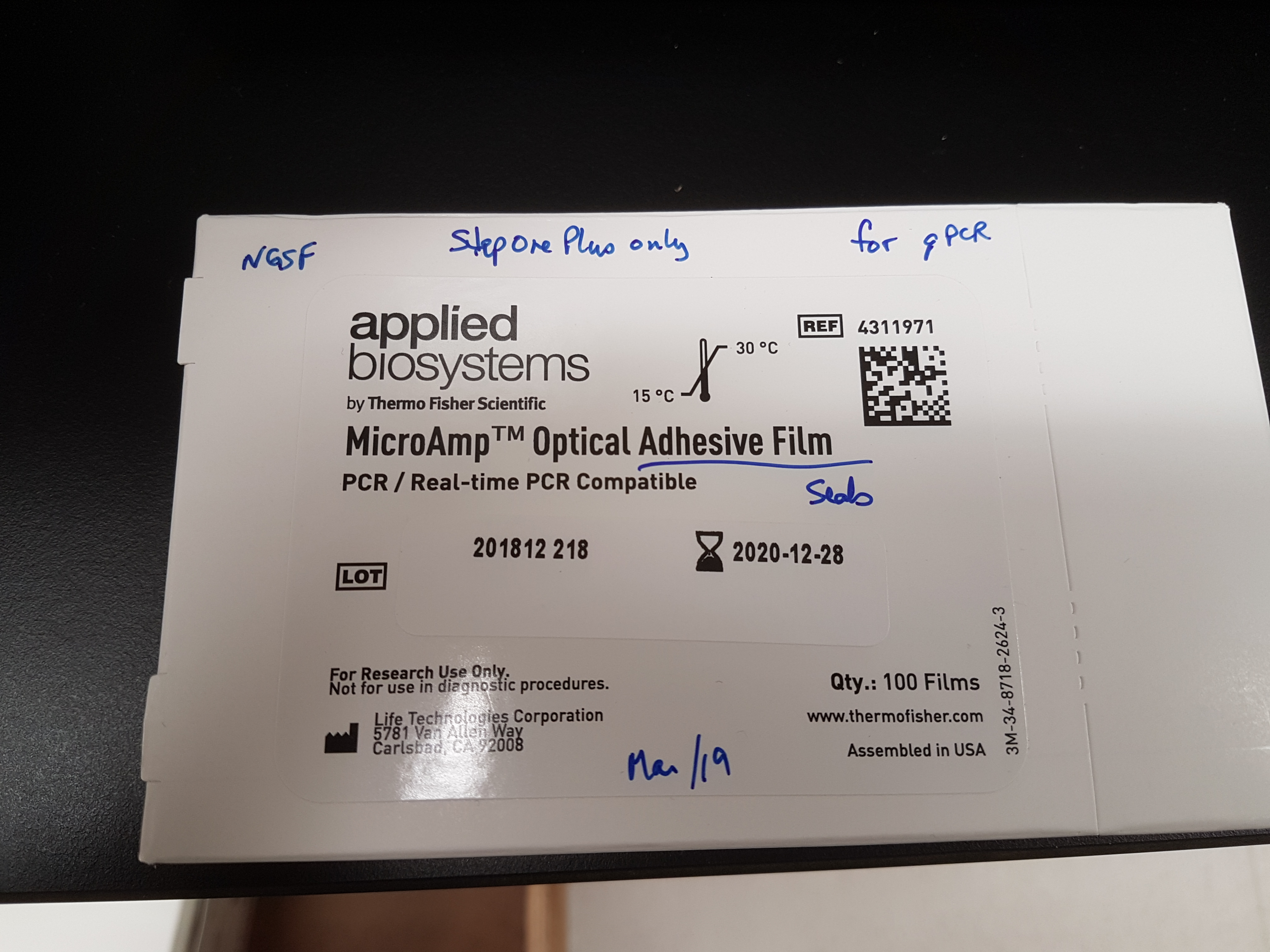
Real-Time PCR
Applied Biosystems StepOne Plus
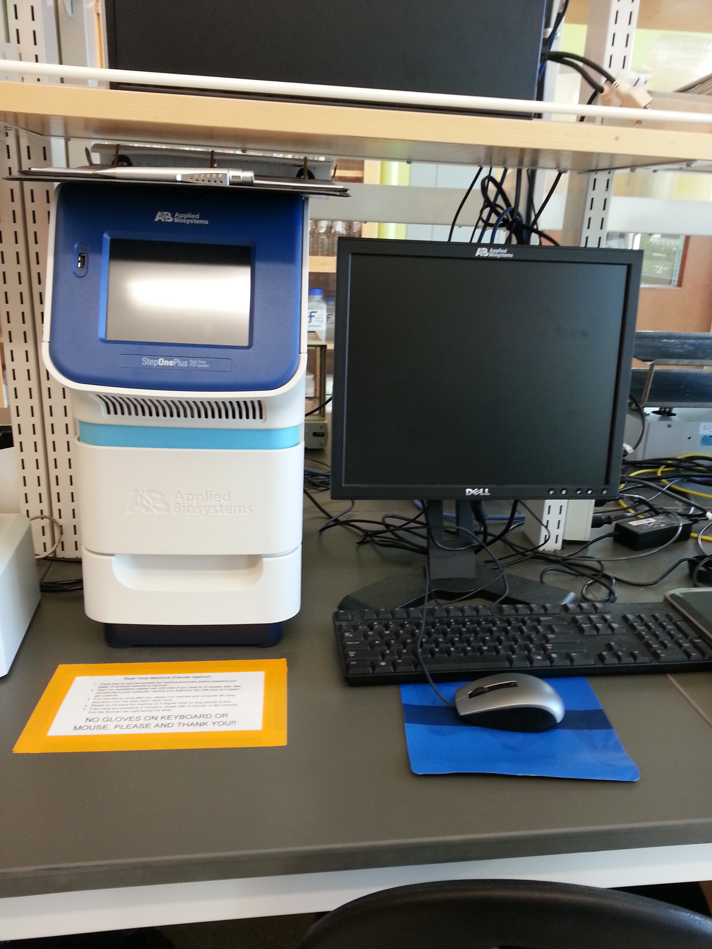
- View Booking Calendar here
- quantitative mRNA expression level measurements
- Users must purchase the appropriate reagents that are compatible with this machine, specifically these exact 96-well plates and seals:
Purchased with funds from the SCA ![]()
Speed Vac
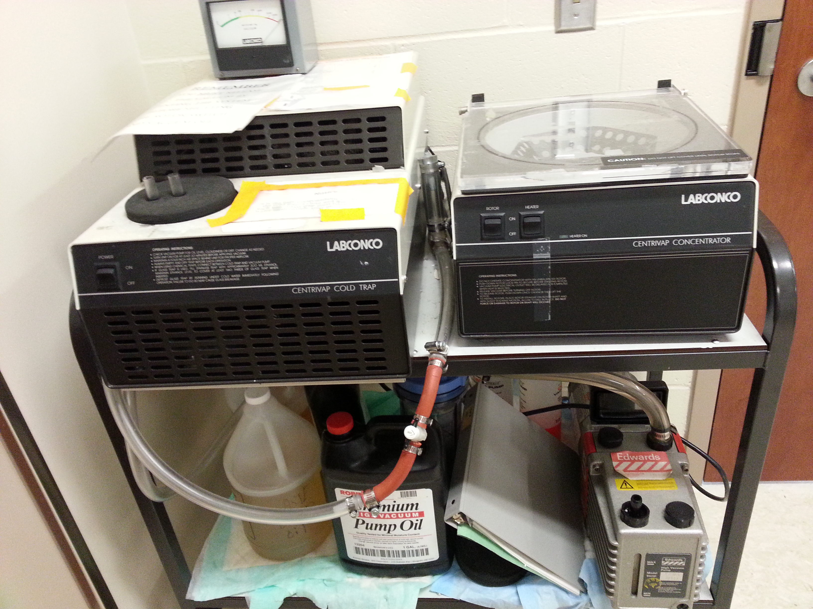
- to dry samples under vacuum in 1.5 mL Eppendorf sized tubes
Ultracentrifuge
Optima L-100 XP Ultracentrifuge
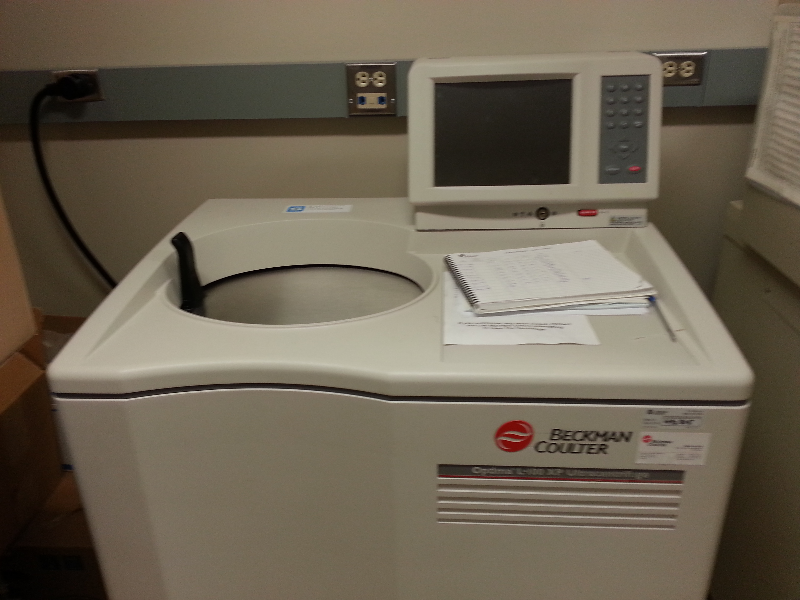
- very high speed centrifugation, under vacuum
Purchased and maintained with funds from the SCA ![]()
xCelligence
RTCA DP Model from ACEA Biosciences
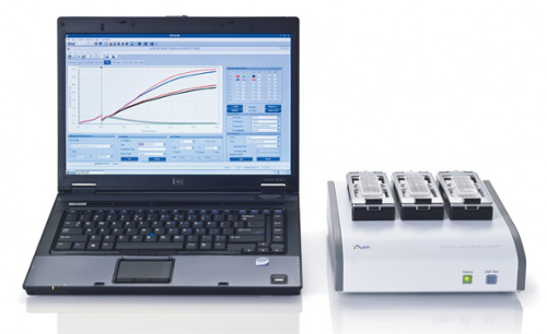
- Live cell real time analyses with multiple measurements over hours and days
- Equipment resides in CO2 incubator
- Cell proliferation measurements
- Cell cytotoxicity measurements (e.g. IC50 determinations)
- Cell migration/invasion measurements
- Morphology change measurements
- specialized 16 well plates with electrodes, that collects data for each of 3 plates independently
- different users can use one of the 3 cradles with independent data collection parameters
- each of the 3 cradles holding the plates must be reserved/booked ahead of time since expts run over multiple days
- measures cell impedance with no dyes or additives needed
- cells can be recovered and analyzed further (e.g. immunoblot or immunofluorescence)
Purchased with funds from the SCA ![]()