

About This Research Area
Working with radiologists and clinical collaborators, we build deep learning systems that read medical images and biomedical signals — and we test them against the specialists they are meant to support.
Early detection of ankylosing spondylitis
About the Project
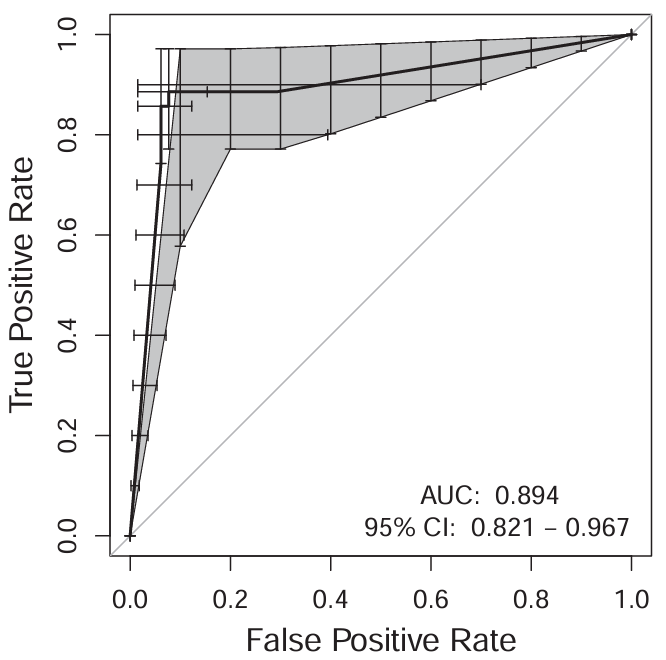
Ankylosing spondylitis is an inflammatory arthritis of the spine and sacroiliac joints that is difficult to catch early, when treatment does most good.
- A YOLOv5 detector locates the sacroiliac joints across 2,025 joints from 1,042 patient frames
- A reduced ResNet50 then grades erosion: 91% accuracy, 100% sensitivity on early-stage cases
- A parallel MRI system outperforms a radiologist with ten years' experience by 13% accuracy
Castro-Zunti et al., Machine Intelligence Research, 2025; Journal of Imaging Informatics in Medicine, 2025.
Rib tracking and fracture classification
About the Project
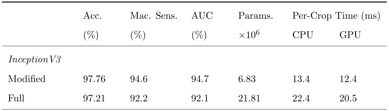
Ribs are the most commonly fractured bones, and finding fractures across a CT stack is slow, repetitive work.
- A U-Net segments the ribs
- Custom multi-object tracking follows each rib through the scan
- Regions are classified as acute fracture, healed fracture or normal
- Validated over 1,000 fracture and 1,000 control scans
RibFractureSys. Castro-Zunti, Li, Vardhan, Choi, Jin and Ko, Computerized Medical Imaging and Graphics, 2024.
COVID-19 detection in chest X-rays
About the Project
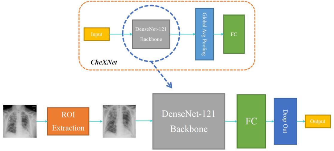
COVID-CXNet detects COVID-19 in frontal chest radiographs, with safeguards against the model reaching the right answer for the wrong reason.
- A U-Net first segments the lungs so the model cannot cheat on irrelevant image features
- A DenseNet-121 backbone performs the classification
- Grad-CAM visualises what drove each decision
COVID-CXNet, Multimedia Tools and Applications, 2022.
Retinal blood vessel segmentation
About the Project
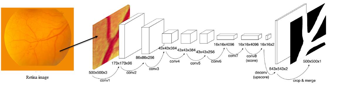
The retina is the one place blood vessels can be photographed non-invasively, which makes it unusually valuable for screening. We segment the vessel tree with a fully convolutional network using transfer learning.
Breast cancer in automated breast ultrasound
About the Project
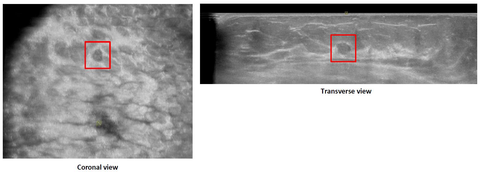
Automated breast ultrasound is non-radioactive and used as supplemental screening for dense breast tissue, where mammography is least reliable.
- A multi-view CNN combines coronal and transverse views of each lesion
- We studied how its performance compares with radiologists at different levels of experience
Choi et al., Ultraschall in der Medizin - European Journal of Ultrasound, 2025.
Dental caries from panoramic X-rays
About the Project
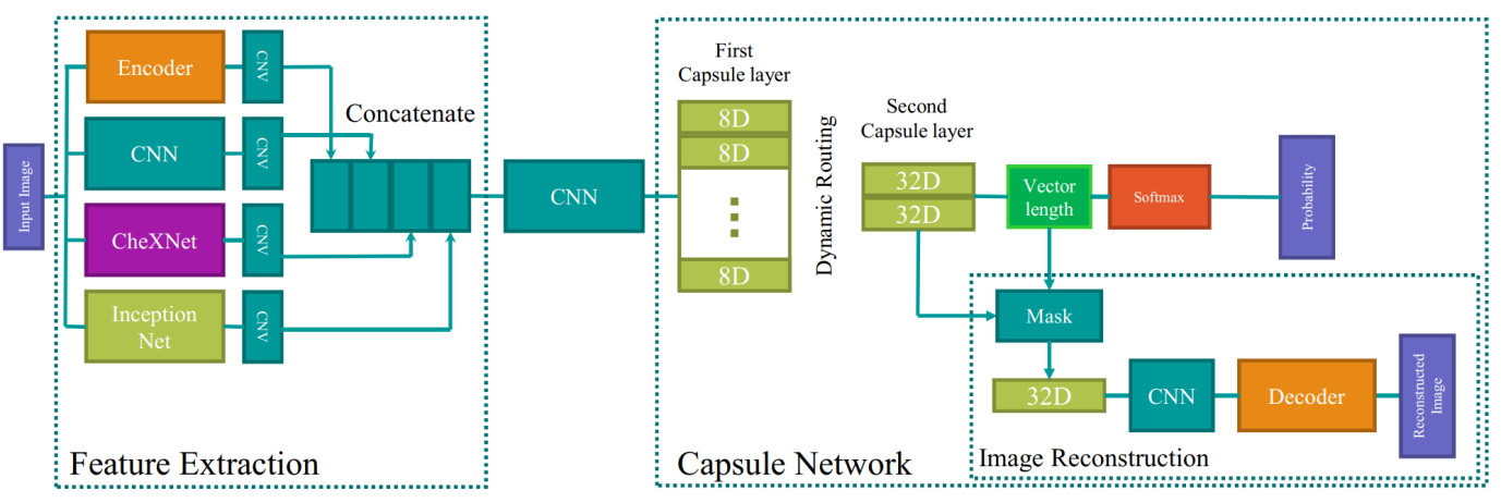
Panoramic dental X-rays are noisy and hard to interpret. PaXNet segments each tooth, then classifies caries.
- CNN feature extraction combined with a capsule network
- An image reconstruction decoder regularises the representation
PaXNet, Multimedia Tools and Applications, 2023.
Biomedical signals
About the Project
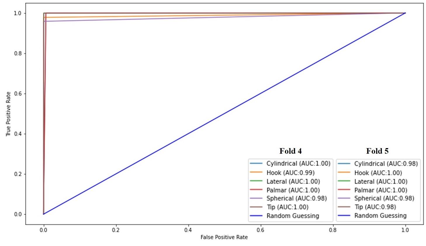
Beyond images, we work on the signals the body produces.
- An LSTM architecture classifying six grasp types from surface electromyography at 99.1% accuracy
- EEG-based biometric authentication through motor imagery
- Transfer learning with self-distillation for detecting schizophrenia from single-channel EEG
Mostafavi et al., Physical and Engineering Sciences in Medicine, 2025.
Also in this area
Further work
- Lung nodule classification on CT (CTLungNet, ten times faster per slice than comparable systems)
- Anterior mediastinal lesion segmentation and diagnosis
- Gout classification from dual-energy CT
- Optimal viewpoint selection for angiography
- Deep learning age estimation from chest CT
- Synthetic contrast-enhanced CT and MRI generated from non-contrast scans