

Dr. Riel Castro-Zunti PhD
Electrical and Computer EngineeringPostdoctoral Fellow in the Ko Lab. His research applies deep learning to medical image analysis, including automated rib-fracture diagnosis and the early detection of disease from radiological images.
Research
Riel applies deep learning to medical image analysis, working with radiologists on systems that catch disease earlier and take repetitive reading work off clinicians.
Publications
Early diagnosis of ankylosing spondylitis
R. Castro-Zunti, E. H. Park, A. Satsangi, Y. Choi, G. Y. Jin, H. S. Chae, S.-B. Ko. Machine Intelligence Research, 2025; and Journal of Imaging Informatics in Medicine, 38(6), 3665-3683, 2025.
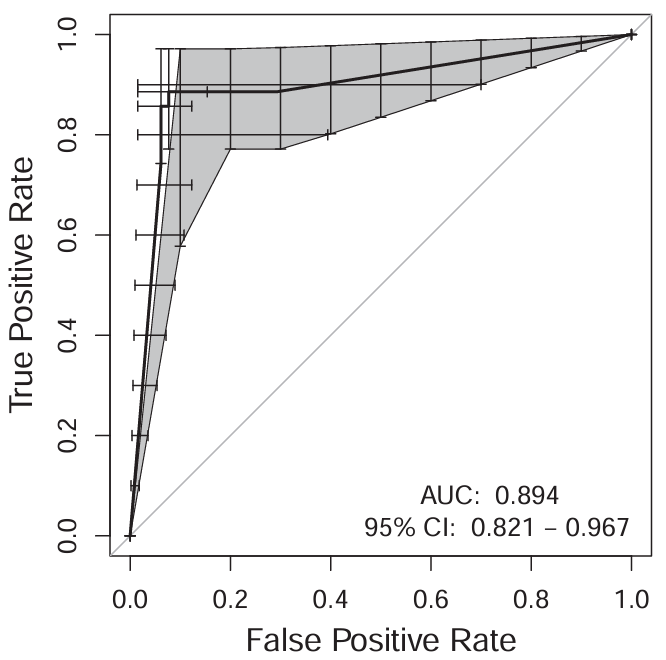
- A YOLOv5 detector locates the sacroiliac joints, then a classifier grades erosion
- 2,025 joints from 1,042 patient frames; 91% accuracy with 100% sensitivity on early-stage cases
- A parallel MRI system outperforms a radiologist with ten years' experience by 13% accuracy
RibFractureSys: rib tracking and fracture classification
R. Castro-Zunti, K. Li, A. Vardhan, Y. Choi, G. Y. Jin, S.-B. Ko. Computerized Medical Imaging and Graphics, 117, 102429, 2024.
- A U-Net segments the ribs and custom multi-object tracking follows each one through the CT stack
- Regions are classified as acute fracture, healed fracture or normal
- Validated over 1,000 fracture and 1,000 control scans
Reducing false positives in gout diagnosis
R. Castro-Zunti, Y. Choi, Y. Choi, H. S. Chae, G. Y. Jin, E. H. Park, S.-B. Ko. Journal of Imaging Informatics in Medicine, 2025.
- A machine vision pipeline identifies and classifies crystal tophi in dual-energy CT
Also
- Tumour-feeding vessel visualisation in cone-beam CT hepatic arteriography (Clinical Radiology, 2026)
- HAZMAT placard detection at highway check stops using synthetic images and YOLOv5-Small (IEEE ISCAS, 2025)